Digital X Ray
Digital radiography is a form of X-ray imaging, where digital X-ray sensors are used instead of traditional photographic film.
Advantages include time efficiency through bypassing chemical processing and the ability to digitally transfer and enhance images. Also less radiation can be used to produce an image of similar contrast to conventional radiography.
Instead of X-ray film, digital radiography uses a digital image capture device. This gives advantages of immediate image preview and availability; elimination of costly film processing steps; a wider dynamic range, which makes it more forgiving for over- and under-exposure; as well as the ability to apply special image processing techniques that enhance overall display of the image.
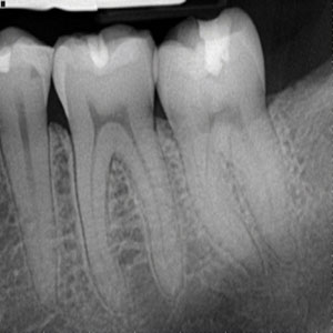
The radiological examinations in dentistry may be classified into intraoral – where the film or sensor is placed in the mouth, the purpose being to focus on a small region of the oral-maxillofacial region and extraoral, where the film or sensor is placed outside the mouth aiming to visualize the entire oral maxillofacial region.
Extraoral imaging is further divided into orthopantomogram, showing a section, curved following more or less mandible shape, of the whole maxillofacial block and cephalometric analysis showing a projection, as parallel as possible, of the whole skull.
Digital radiography in dentistry provides the clinician with the ability to store their images on a computer. This provides two key advantages over film in the form of full screen images that can be enhanced and zoomed in on, aiding diagnostics and providing easier patient communication, as well as allowing dental offices to communicate images electronically, allowing for simpler referrals and, where applicable, easier insurance claim submission.